When a wrist fracture doesn’t heal properly, it can leave behind more than just discomfort. It can limit mobility, cause long-term pain, and even change the appearance of the wrist. These improperly healed breaks, called malunions, often require surgical correction to restore the wrist’s natural function and shape.
At Elite Sports Medicine + Orthopedics, orthopedic hand surgeon Dr. Tom Dovan is using an innovative technique to help patients reclaim their quality of life: 3D-printed osteotomy for distal radius malunion. This advanced surgical solution combines orthopedic skill with custom-designed technology to create a more accurate and personalized treatment.
What Is a Distal Radius Malunion?
The distal radius is the part of the forearm bone closest to the wrist. It’s one of the most commonly fractured bones in the body, especially from falls. In some cases, a wrist fracture can heal out of alignment—either due to poor positioning, instability during healing, or removal of a cast too soon. When this happens, the wrist may not move the way it should, and it may cause pain or deformity that interferes with daily life.
This is especially problematic for people whose work or passion relies on precision wrist motion—such as athletes, musicians, or dancers.
The Surgical Innovation: 3D-Printed Osteotomy
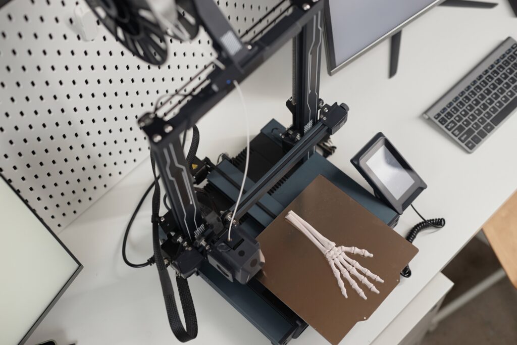
Traditionally, osteotomies (surgical bone realignment procedures) are performed using 2D X-rays and the surgeon’s expert judgment. But now, 3D-printing technology is changing the game, giving surgeons greater accuracy and control, tailored to each patient’s exact anatomy.
Here’s how Dr. Dovan’s approach works:
- CT Scanning and Digital Modeling
Both wrists are scanned using CT imaging to create 3D models of the bones. This allows the surgical team to compare the injured wrist with the healthy one and precisely identify the areas that need correction.
- Virtual Surgical Planning
Using advanced planning software, the team maps out the osteotomy in detail, calculating the exact cuts needed to realign the bone and restore proper function.
- Custom 3D-Printed Surgical Guides
Next, custom tools are 3D-printed based on the plan. These surgical guides are made specifically for the patient and help guide each cut and screw placement during surgery.
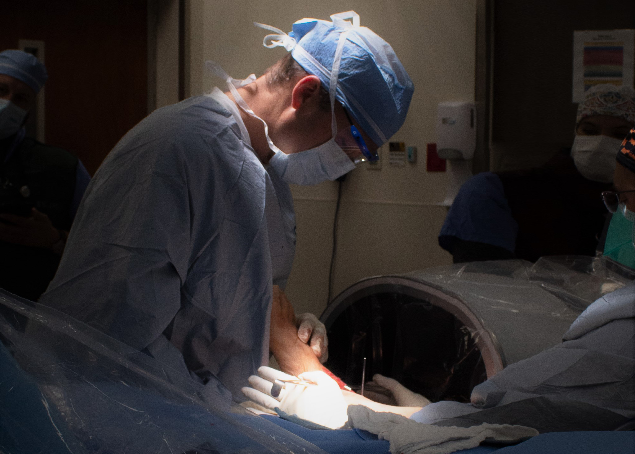
- Surgical Execution
During the procedure, Dr. Dovan uses the guides to make precise bone cuts and place hardware with accuracy that would be difficult to achieve using traditional methods. The goal is to replicate the healthy wrist’s anatomy as closely as possible.
Why 3D Technology Makes a Difference
This approach offers several advantages compared to conventional methods:
- Tailored Accuracy: Because the surgical plan is based on the patient’s own anatomy, the result is more precise and individualized.
- Reduced Error: Custom guides help ensure that everything is done exactly as planned.
- Improved Recovery: Patients often regain better range of motion and experience less pain when alignment is restored correctly1.
The use of 3D printing in orthopedic surgery reflects a growing trend toward personalized, tech-driven care—one that combines cutting-edge tools with surgical expertise.
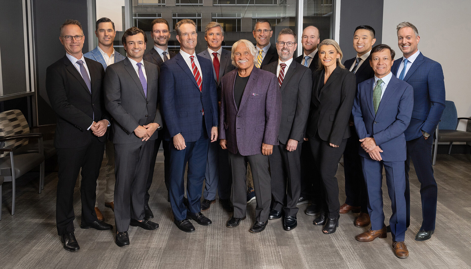
Leading the Way at Elite
Dr. Tom Dovan is one of the few surgeons in the region performing this highly specialized technique. His work at Elite Sports Medicine + Orthopedics is helping redefine what’s possible for patients dealing with complex wrist injuries.
Whether you’re a professional athlete or someone who simply wants to move without pain, 3D-printed osteotomy offers a new level of hope—and healing.
To schedule a consultation with Dr. Dovan, book online or call our office today.
Footnotes
- Giesen, E. B., et al. “Patient-specific 3D planned corrective osteotomies in malunited distal radius fractures.” Journal of Orthopaedic Surgery and Research, 2019. https://doi.org/10.1186/s13018-019-1212-5 ↩

Author: Nick Flory
Related Articles
We are proud to share that ten of our physicians at Elite Sports Medicine and Orthopedics have been named Nashville Top Doctors by Castle Connolly [...]
Thumb arthritis can make everyday tasks like opening jars, turning keys, gripping objects, and pinching painful and frustrating. For patients with advanced thumb arthritis who [...]

Shoulder pain is one of the most common reasons patients visit a sports medicine specialist or orthopedic doctor. It affects athletes, active adults, and [...]

Joint pain is one of the most common health complaints across all age groups. Most everyday aches improve with rest, ice, or over-the-counter medication—but sometimes [...]
When athletes think about improving performance, the focus often lands on strength training, conditioning, and skill development. One critical factor is often overlooked: foot health. [...]

When we talk about sports injuries, we often picture athletes sprinting down a field or pushing through an intense workout. But you don’t have to [...]
In April 2024, Suzanne’s life changed almost instantly. What started as a normal Sunday evening turned into a frightening medical emergency, and her experience shows [...]

Staying active as we age isn’t just about fitness. It’s about staying independent, energetic, and living life on your terms. As more adults embrace active [...]
Knee pain after a sudden twist or landing? You might be facing more than a simple strain. ACL injuries are among the most common and [...]

Around one in every two adults in the United States live with a musculoskeletal condition, making it the leading cause of disability nationwide. With so many [...]